Information About Cicatricial Alopecia or Scarring Hair Loss

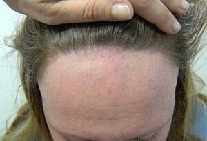
Frontal Fibrosing Alopecia

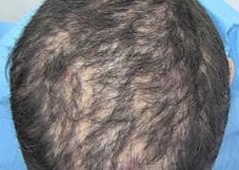
Folliculitis Decalvans

Folliculitis Decalvans

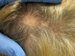
Lichen Planopilaris

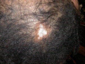
Discoid Lupus Erythematosus
Cicatricial Alopecia - Dirk M. Elston, MD & Elise Olsen, MD
See FAQs at the bottom of the page.
Cicatricial (scarring) alopecia (hair loss) is the term used for a group of disorders that cause permanent hair loss. During the active, evolving stage of hair loss, patches of alopecia commonly appear red and inflamed at the base of the hair shaft. Sometimes crops of pustules are noted. Some types of cicatricial alopecia destroy the hairs deep within the scalp, without inflammation visible on the skin surface. While some types of cicatricial alopecia result in rapid hair loss, slow progression of hair loss is more common.
A skin biopsy is generally required to establish the diagnosis, and to guide treatment. The biopsy punch is an instrument that removes a plug of skin about the size and shape of a pencil eraser. The biopsy is performed after an injection of local anesthetic to numb the skin. After the skin biopsy is removed, the biopsy site is closed with stitches or filled with a plug of special material that stops the bleeding. Sometimes a single biopsy specimen can establish the diagnosis, but usually more than one specimen is required. Your doctor will try to limit the number of biopsy specimens, but it is generally best to have more than one performed early on in your evaluation, so that an accurate diagnosis can be established and appropriate treatment started.
Some types of hair loss are best diagnosed under the microscope based on slices of the specimen cut vertically from the skin surface down to the deep fat (vertical sections). Other types of hair loss are best diagnosed by horizontal sections cut sideways through the specimen (horizontal sections). Each of these types of examination requires a separate biopsy specimen. Biopsies are best done of active, inflamed sites on the scalp which still have remaining hair. A biopsy of an older scarred area may be helpful to predict the likelihood of regrowth of hair, and to help establish the diagnosis by evaluating the pattern of scar formation. If certain types of cicatricial alopecia are suspected, your doctor may send a biopsy specimen for additional special tests including direct immunofluorescence, and special stains for bacteria, fungi and elastic tissue. In some infectious disorders that can cause cicatricial alopecia, a biopsy must be sent for tissue culture.
At the 2001 AHRS Workshop on Cicatricial Alopecia held at Duke University Medical Center, a useful classification system for cicatricial alopecia was developed, emphasizing the microscopic (histological) findings on a representative biopsy of an area of active loss. This classification divides cicatricial alopecia into hair loss caused by inflammatory cells called lymphocytes versus hair loss caused by inflammatory cells called neutrophils. The new classification will help identify patients who may be appropriate candidates for studies of new treatments since it is likely that medications will not work on both lymphocytic and neutrophilic predominant cases of cicatricial alopecia. Even with the new histological classification, some cases of hair loss remain unclassifiable.
Treatment for cicatricial alopecia remains poor. Once the hair is destroyed, the hair loss becomes permanent. It is primarily the hair at the periphery of the hair loss and/or the islands of remaining hair that are at risk of being destroyed that is the main focus of treatment. The main goals of treatment for cicatricial alopecia are to prevent further hair loss and to eradicate or at least lessen the redness, scale and itching associated with the process. There are no current FDA approved treatments for cicatricial alopecia although the AHRS has been working to initiate interest in this area among industry sponsors. All treatment now for cicatricial alopecia is strictly based on the experience of the prescribing physician or anecdotal reports as there has never been any multicenter clinical trial in this area.
MAJOR TYPES OF CICATRICIAL ALOPECIA CAUSED BY LYMPHOCYTES:
MAJOR TYPES OF CICATRICIAL ALOPECIA CAUSED BY NEUTROPHILS:
SUMMARY:
There are many other less common types of cicatricial alopecia. A careful physical examination, scalp biopsies and blood tests can be helpful in order to establish the correct diagnosis and to suggest the most appropriate treatment for the hair loss. Many patients do not respond to the first treatment they receive and often the condition relapses when treatment is stopped. The new AHRS classification system for cicatricial alopecia was designed to help group patients who might respond to promising new treatments. The AHRS multicenter study of CCCA in African American women may hopefully lead to an explanation and effective treatment for this condition. If you have cicatricial alopecia, your dermatologist can help guide you through the array of off-label and experimental treatments that are available.
References:
Cicatricial Alopecia Frequently Asked Questions:
The following are frequently asked questions on cicatricial alopecia. The information provided is not meant to be a substitute for the information obtained at an evaluation and by discussion with a physician, but merely to encourage understanding of this condition. No questions regarding individual scenarios will be answered by the AHRS. No changes in treatment should be undertaken by a patient without discussion first with the patient's physician.
In all forms of cicatricial alopecia, fibrous tissue replaces the hair follicles. In most conditions, the inflammatory cells destroy all the appendages (hair, oil and sweat glands) in an area of the scalp and an area with hair is replaced by hairless skin that appears "slick", without the usual visible pores, and may be slightly depressed on palpation. It is "like a scar" but does not necessarily have the appearance of a scar that occurs after trauma ie it may not look very different from normal skin to most people. It is not the result of a break in the skin being closed by scar tissue. In cicatricial aloplecia, the scar is mostly underneath the surface where there is gradual thickening of the fibrous tissue.
The hair loss you describe fits the description of central centrifugal cicatricial alopecia (CCCA). As a group, patients with this disorder have similar clinical and biopsy findings, however the cause of the disorder remains unknown. No treatment has been proved to be effective although topical corticosteroids and oral antibiotics are sometimes sometimes help stabilize the condition and prevent it from getting worse. Because it has been suggested that the disorder may be related to hair grooming practices, many physicians recommend avoiding chemical treatments and excessive hair tension. Research into this condition continues, including work currently being done by the NAHRS. Once lost, the hair rarely regrows so the emphasis must be on preventing further loss.
Most patients with chronic cutaneous lupus erythematosus of the scalp do not have systemic lupus erythematosus, and will never develop internal problems related to lupus. A physical examination together with blood and urine tests can be used to determine who is likely to have internal problems with their lupus. Depending on the severity of the skin lupus, you may require courses of oral treatment in addition to topical treatments. Cicatricial alopecia caused by lupus may actually show some hair regrowth with treatment which is different from other types of cicatricial alopecia where prevention of further loss and not regrowth is the hoped for result of effective therapy.
The findings you describe suggest a diagnosis of Dissecting Cellulitis of the scalp. Although bacteria play a role in this disorder, it is not simply an infection of the skin. The disorder resembles severe cystic acne of the scalp. Long term use of antibiotics, together with drainage and injection of individual lesions may be effective. Many patients need a combination of treatments. Vitamin A derivatives, also called retinoids, have shown some success with this problem.
First, it is important to establish the correct diagnosis. If you have not had scalp biopsies, or if they were inconclusive, additional biopsies are the best means of establishing the diagnosis. Tests such as direct immunofluorescence, tissue culture and special stains may be helpful in establishing the diagnosis. Some conditions that can mimic LPP include lupus erythematosus and folliculitis decalvans (FD). Biopsy will often distinguish these entities. If you experience periodic crops of pustules on the scalp, the diagnosis is more likely to be FD. FD requires long-term treatment with antibiotics. The antibiotics used to treat FD are often different from those used to treat LPP.
Having said this, the hair loss in the pattern you describe is often caused by lichen planopilaris (LPP). LPP can be difficult to treat, and may not respond to antibiotics and topical steroids. Other treatments such as oral or intralesional corticosteroids (cortisone-type drugs), retinoids, antimalaria pills such as plaquenil or thalidomide may be useful in some cases.
Cicatricial Alopecia Path Project
CPATHOLOGY EVALUATION FOR CICATRICIAL ALOPECIA (Version 2)
Purpose: This form was created during the North American Hair Research Society (AHRS) – sponsored workshop on cicatricial alopecia (Feb 10 and 11, 2001). The intent was to create a standardized template for recording pathologic findings in patients with cicatricial alopecia. The original form (version 1) has already been published (J Amer Acad Dermatology 2003; 48: 103 – 110).
An electronic version of the form (version 2) was created as part of a AHRS – sponsored study of the utility of the form in actual use by dermatopathologists experienced in the pathologic evaluation of alopecia. This version is now available to physicians who wish to utilize or test the form while recording findings from their own pathologic specimens.
(The electronic version is MS Excel format and contains macros. Security settings within Excel will need to be set to medium or lower to open the file.) PDF VERSION HERE (text only)
Questions or comments regarding the form can be directed to: lsperling@usuhs.mil
